



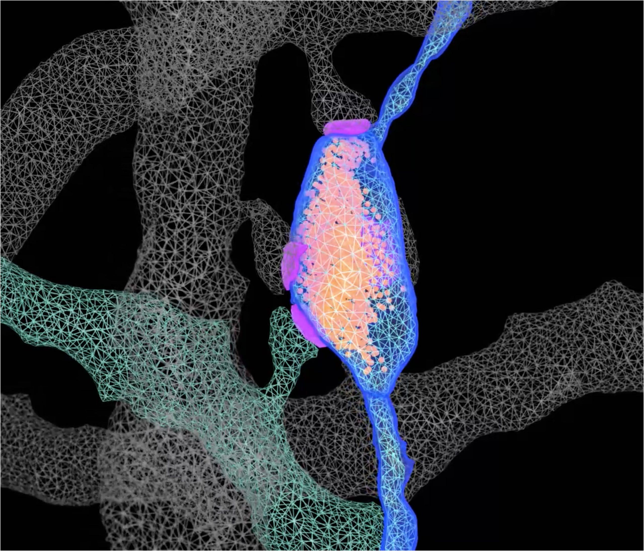
An engram, a key characteristic of long-term memory, has been identified by Scripps Research scientists and their collaborators using cutting-edge genetic tools, 3D electron microscopy, and artificial intelligence. Published in Science on March 20, 2025, their findings offer new insights that could lead to improved treatments for memory loss and other cognitive impairments linked to aging and neurodegenerative diseases.
“Our work leverages recent technological developments across multiple fields,” says Marco Uytiepo, the study’s principal author and a Scripps Research graduate student. “We employed high-resolution 3D imaging to show the intricate architecture of brain circuits that retain memory traces in unprecedented detail. Because analyzing these photos using traditional computer programs could take years, we relied significantly on AI algorithms to speed up data processing by several orders of magnitude.”
Uytiepo and his team concentrated on the hippocampus, a brain area critical for learning and memory in both animals and humans. Using mouse models, they tagged and identified neurons that were active during a specific learning challenge. They then recreated the synaptic connections between these neurons, where communication happens, with nanometer-scale detail.
“We hoped to uncover something interesting because no similar approaches had previously been implemented,” says Anton Maximov, a neuroscience professor and the study’s senior author. “What we did not anticipate was that our findings would challenge two long-held dogmas,” according to the authors.
Displacing Conventional Theories of Memory Formation
Chemical signals at neuronal synapses are normally conveyed from a single nerve terminal—a swelling portion of an axon loaded with vesicles that leak these signals—to a single postsynaptic location on a receiving cell’s dendrite. Many prior research (with lower-resolution optical imaging technologies) have revealed that learning necessitates a significant increase in synapse number.
Maximov’s team discovered that this is not always the case, since the total number and layout of isolated synapses remained constant following memory formation. Instead, neurons assigned to an engram increased their connectivity via multi-synaptic boutons (MSBs), which are specialized axonal terminals that signal to up to six separate dendrites instead of just one.
Unexpected Cellular Changes and Network Behavior
Second, Maximov’s team discovered that engram neurons in nearby hippocampus areas do not link preferentially, contrary to popular belief in the field. Instead, expanding their network via MSBs resulted in the recruitment of neurons that were not active during learning. Furthermore, the researchers discovered that engram neurons showed fine-scale modifications in the architecture of their individual synapses, including changes in intracellular organelles like mitochondria and the smooth endoplasmic reticulum. Furthermore, these neurons had stronger connections with astrocytes, which regulate synaptic activity and offer metabolic support.
Researchers are now investigating if comparable mechanisms exist in other brain circuitry and whether their failure contributes to memory loss. Furthermore, MSBs have been identified as promising therapeutic targets.
“We are excited about the possibility of targeting MSBs with drugs to develop new and effective treatments for memory disorders,” according to Maximov. “However, reaching this goal will necessitate developing new methods to examine the molecular composition of MSBs, which is currently untapped. We are already making headway in this area, but much more work remains.”
As part of this work, the researchers are constantly refining their AI pipelines to improve the efficiency and accuracy of interpreting large-scale imaging data.
This work was undertaken in partnership with UC San Diego’s National Center for Microscopy and Imaging Research (NCMIR), which is led by Distinguished Professor of Neurosciences Mark H. Ellisman. NCMIR, an NIH BRAIN Initiative National Resource for Technology Integration and Dissemination, offers cutting-edge imaging techniques to promote neuroscience research.
“We consider ourselves extremely fortunate to have partnered with Mark and his team,” Maximov says. “Our success was greatly aided by their extensive knowledge, technical proficiency, and access to cutting-edge microscopes.”

